


Coksartrosis- This is the arthrosis of the hip joint.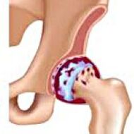 It develops gradually, for several years, subject to progression, can be both a side and a double side.It is accompanied by the pain and restriction of movements in the articulation.In the following stages the atrophy of the hip muscles and the shortening of the limb are observed.The diagnosis is established on the basis of the clinical symptoms and the results of the X -ray.In the early stages of coxartrosis, conservative treatment.With the destruction of the joint, especially in young and middle patients, surgery (endoprothetic) is indicated.
General information
Coksartrosis (osteoarthritis or arthrosis deforming of the hip joint) is a degenerative dyeing disease.Usually it develops at the age of 40 and more.It can be the result of various injuries and joint diseases.Sometimes it occurs for no apparent reason.Coksartrosis is characterized by a gradual progressive course.Conservative treatment methods are used in the early stages.In the subsequent stages, the joint function can only be restored operational.
In orthopedics and traumatology, coxartrosis is one of the most common arthrosis.The high frequency of its development is due to a significant load on the hip joint and the widespread prevalence of congenital dysplasia - joint dysplasia.Women suffer from coksartrosis a little more often than men.
The causes of Coksartrosis
The primaries (deriving from unknown reasons) and secondary (developed following other diseases) are distinguished, the arthrosis of the hip joint.
Secondary Coksartrosis can be the result of the following diseases:
- Hip dysplasia.
- Innate displosure of the thigh.
- Pertes diseases.
- Aseptic necrosis of the head of the thigh.
- Infectious lesions and inflammatory processes (for example the arthritis of the hip joint).
- Injuries (traumatic dislocations, hip neck fractures, pelvic fractures).
Coksartrosis can be a side or double side.With primary coxartrosis, there is often a concomitant injury of the spine (osteochondrosis) and knee joint (gonartrosis).
Risk factors
Among the factors that increase the probability of the development of coxartrosis include:
- CONSTERNERS CONSTITUTE ON THEMore often observed in athletes in people with excess body weight.
- Circulatory disorders, hormonal changes, metabolic disorders.
- Vertebral column pathology (kyphosis, scoliosis) or stop (flat feet).
- Elderly and senile age.
- A sedentary lifestyle.
Coksartrosis itself is not inherited.However, some characteristics (metabolic disorders, structural characteristics of skeleton and weakness of cartilage) can be inherited from the child by the parents.Therefore, in the presence of blood relatives suffering from coxarthrosis, the probability of the onset of the disease has slightly increased.
PataNatomy
The hip joint is made up of two bones: ileo and femoral.The head of the thigh is articulated with the acetabulum of the iliac bone, forming a peculiar "zipper".During the movements, the acetabulum remains motionless and the head of the femur moves in various directions, guaranteeing flexion, extension, kidnapping, bearing and rotational hips.
During the movements, the joint surfaces of the bones without obstacles to each other, thanks to the cartilage ialina smooth, elastic and resistant that covers the cavity of the swivel cavity and the head of the thigh.In addition, cartilage IALINA performs a shock absorption function and is involved in the redistribution of the load during movement and walking.
In the joint cavity there is a small amount of joint liquid, which plays the role of lubrication and provides nutrition of the cartilage IALINA.The joint is surrounded by a dense and strong capsule.Above the capsule there are great femoral and buttock muscles, which provide movements in the joint and, together with the cartilage IALINA, are also shock absorbers that protect the articulation from injuries with unreacked movements.
With coxartrosis, the joint liquid becomes more often and more viscous.The surface of the cartilage IALINA dries, loses the softness, covered by cracks.Due to the roughness that arose, the cartilage during the movements is constantly injured one towards the other, which causes their thinning and aggravates pathological changes in the articulation.As coxartrosis advances, the bones begin to deform, "adapting" to the increase in pressure.The metabolism in the joint is deteriorating.In the subsequent stages of coxartrosis, a serious atrophy of the muscles of the painful limb is observed.
Symptoms of coxartrosis
The main symptoms of the disease include pain in the articulation of the joint, of the inguinal region, thigh and knee.In addition, with cokes arthrosis, rigidity of the movements and rigidity of the articulation, disorder of the pace, lameness, atrophy of the hip muscles and shortening of the limb on the side of the lesion.A characteristic characteristic of Coksartrosis is a restriction of kidnapping (for example, the patient is difficult when trying to sit on a chair).The presence of some signs and their gravity depends on the stadium of Coxartrosis.The first and most constant symptom is pain.
TOCoksartrosis of the 1st degreePatients complain of periodic pain, which occurs after physical activity (running or prolonged walk).The pain is located in the joint, less often in the thigh or knee.After rest, it usually disappears.The pace for the 1st degree coxartrosis is not broken, the movements are preserved in full, there is no muscle atrophy.
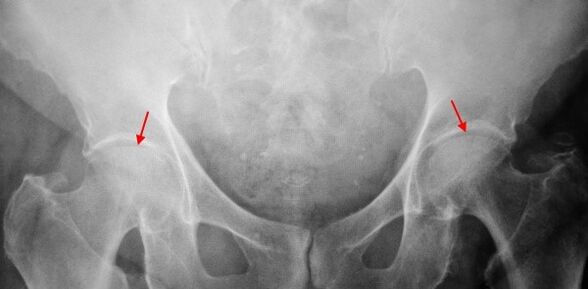
On the ray X of the patient suffering from 1st degree coxartrosis, mild changes are determined: a moderate irregular narrowing of the joint gap, as well as the bone growth around the external or internal edge of the acetabulum in the absence of changes from the head and neck of the femur.
TOCoksartrosis 2 degreesPain becomes more intense, it often appears at rest, it radiates in the thigh and groin.After a significant physical activity, the patient with Coksartrosis begins to limp.The volume of movements in the joint decreases: the abduction and internal rotation of the thigh are limited.
In the X -ray images for the 2nd degree coxartrosis, a significant irregular narrowing of the joint gap (more than half of the normal height) is determined.The head of the femur is somehow moved upwards, deformed and increases in size and its contours become irregular.Bone growth with this degree of coxartrosis appear not only on the interior, but also on the outer edge of the acetabulum and go out of the cartilage.
TOCoksartrosis 3 degreesPain becomes constant, concern for patients not only during the day, but also at night.Walking is difficult, when it moves, a patient with coksartrosis is forced to use a stick.The volume of movements in the joint is abruptly limited, the buttock muscles of the buttock, the lower sides and legs are atrophied.The weakness of the thigh removal muscles becomes the cause of the deviation of the pelvis in the front plane and shortening the limb on the sore side.In order to compensate the shortening, a patient who suffers from coksartrosis, when walking, incurs the body in the painful direction.For this reason, the center of gravity moves, the load on the sore joint increases sharply.
A strong narrowing of the joint gap, a pronounced expansion of the head of the thigh and more bone outcrops, are detected on radiographs for coxarthrosis of the 3rd degree.
Diagnostics
The diagnosis of coxartrosis is based on clinical signs and data of further studies, whose main is the radiography.In many cases, X -rays allow you to establish not only the degree of coxartrosis, but also the cause of its occurrence.Therefore, for example, an increase in the corner of the neck-diaphysal, the scenes and flattening of the acetabulum indicate dysplasia and the changes in the form of the proximal part of the femur are indicated that coks arthrosis is a consequence of the perties disease or youth epiphyphysiolisi.On the radiographs of patients with coxartrosis, it is possible to detect changes that indicate injuries.
Like other methods of instrumental diagnosis of coxartrosis, it is possible to use CT and magnetic resonance imaging.The computerized tomography allows you to study the pathological changes in detail by the bone structures in detail and the magnetic resonance imaging imaging offers the opportunity to evaluate the disorders by soft tissues.
Differential diagnosis
First of all, coxartrosis should be differentiated by gonartrosis (osteoarthritis of the knee joint) and osteochondosis of the spine.The atrophy of the muscles, which occurs in 2 and 3 phases of coxartrosis, can cause pain in the knee joint, which are often expressed brighter than pain in the damage area.Therefore, with the patient's complaints about knee pain, a clinical inspection (inspection, palpation, determination of the volume of movements) is the study of the hip joint and if coxarthrosis is suspected, to direct the patient to the X -ray.
The pain for root syndrome (compression of the nerve roots) for osteochondrosis and some other diseases of the spine can imitate pain with coxartrosis.Unlike coxartrosis, when it tightens the roots, the pain suddenly occurs, after a movement without success, an acute turning point, the lifting weights, etc., is located in the buttock and spreads along the back of the thigh.A positive symptom of tension is detected: severe pain when the patient tries to raise a straightened limb, lying on his back.At the same time, the patient freely takes the leg on the side, while in patients with coksartrosis, the kidnapping is limited.It should be borne in mind that osteochondrosis and coksartrosis can be observed simultaneously, therefore, in all cases, an in -depth examination of the patient is necessary.
In addition, cokesartrosis is differentiated with trocantery (booting) - aseptic inflammation in the attack area of the buttock muscles.Unlike coxartrosis, the disease develops quickly, within 1-2 weeks, usually after a significant injury or physical activity.The intensity of the pain is higher than Coksartrosis.There are no limits of movements and shortening of the limb.
In some cases, with an atypical course of the disease or reactive arthritis, symptoms that recall coxartrosis can be observed.Unlike coxartrosis, with these diseases, the peak of pain falls at night.Pain syndrome is very intense, it can decrease when walking.The morning rigidity is characteristic, which occurs immediately after awakening and gradually disappears in a few hours.
Coxartrosis treatment
The treatment of pathology is engaged in traumatologists orthopedists.The choice of treatment methods depends on the symptoms and the disease phase.At 1 and 2 stages of coxartrosis, conservative therapy is performed.During the period of exacerbation of coxartrosis, injection blocks, anti -anti -inflammatory drugs non steridal (pyroxic, indomethacin, diclofenac, ibuprofen, etc.).It should be borne in mind that the drugs of this group are not recommended for a long time, since they can have a negative effect on the internal organs and suppress the ability of the cartilage IALINA to restore.
To restore damaged cartilage for coksartrosis, funds from a group of chondroprotectors (Condroitin sulphate, cartilage extract, etc.) are used.To improve blood circulation and eliminate the spasm of small vessels, vasodilant drugs are prescribed (zinnarisine, nicotine acid, pentoxifillin, xanthinol nicotinato).According to the indications, muscle relaxing (muscle relaxation drugs) are used.
With stubborn pain syndrome, patients suffering from coksartrosis may be prescribed intra -articular injections using hormonal drugs (hydrocortisone, triamcinolone, metumor).The treatment with steroids should be carried out with caution.In addition, with coxartrosis, local products are used: heating ointments that do not have a pronounced therapeutic effect, however, in some cases they relieve muscle spasm and reduce pain due to their "distracted" action.Furthermore, with coxartrosis, physiotherapy procedures are prescribed (bright, ultrasonic therapy, laser treatment, uhf, inductothermia, magnetotherapy), massage, manual therapy and therapeutic gymnastics.
The diet for Coksartrosis does not have an independent therapeutic effect and is used only as a means of reducing weight.The reduction of body weight allows you to reduce the load on the hip joints and, consequently, facilitate the course of Coksartrosis.In order to reduce the load on the joint, the doctor, depending on the degree of coxartrosis, may recommend walking with a stick or crutch.
In the subsequent stages (with 3rd degree coxartrosis), the only effective treatment method is the operation: the replacement of the articulation destroyed with an endoprotesis.Depending on the nature of the lesion, it can be used or replacing only the head of the thigh) or two -Pole (which replaces both the head of the thigh and the swivel cavity).
The Endopropostetic operation for coxartrosis is carried out in a planned way, after a complete examination, under general anesthesia.In the postoperative period, antibiotic therapy is performed.The seams are removed in 10-12 days, after which the patient is prescribed for outpatient treatment.After endoprothetics, rehabilitation measures are necessarily supported.
In 95% of cases, the surgery to replace the articulation with coxartrosis guarantees a complete restoration of the function of the limbs.Patients can work, move actively and even do sports.The average duration of the prosthesis, without prejudice to all the recommendations, is 15-20 years.Subsequently, a second operation is needed to replace a worn endoprotesis.